


Services on Demand
Journal
Article
 English (pdf)
 Article in xml format
 Article references
 Send this article by e-mail
Indicators
-
 Cited by SciELO
 -
 Access statistics
Related links
-
 Similars in
SciELO
Similars in
SciELO
Share

 Permalink
GE-Portuguese Journal of Gastroenterology
Print version ISSN 2341-4545
GE Port J Gastroenterol vol.26 no.5 Lisboa Oct. 2019
https://doi.org/10.1159/000494095
IMAGES IN GASTROENTEROLOGY AND HEPATOLOGY
Capsule Endoscopy in the Diagnosis of Eosinophilic Enteritis
Cápsula endoscópica no diagnóstico de enterite eosinofílica
Mafalda Sousa, Rolando Pinho, Luísa Proença
Gastrenterology Department, Centro Hospitalar de Vila Nova de Gaia e Espinho, Vila Nova de Gaia, Portugal
* Corresponding author.
Keywords: Capsule endoscopy, Eosinophilic enteritis, Nonspecific findings
Palavras-Chave: Cápsula endoscópica, Enterite eosinofílica, Achados inespecíficos
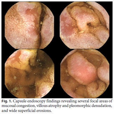
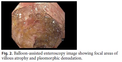
A 45-year-old female patient with allergic rhinitis without medication was referred for evaluation for suspected Crohns disease in 2004. She presented with epigastralgia, dyspepsia, and iron deficiency anemia. Upper digestive endoscopy showed multiple erosions in the 2nd and 3rd duodenal portions; ileocolonoscopy was normal, and capsule endoscopy revealed 10 aphtoid erosions in the duodenum, jejunum, and ileum. Peripheral eosinophilia and elevated serum IgE were absent, and antitransglutaminase and stool for ova and parasites were negative. Surveillance without treatment was recommended, and the patient was lost to follow-up until 2017 when capsule endoscopy was repeated and revealed several focal areas of mucosal congestion, villous atrophy and pleomorphic denudation, and wide superficial erosions in the duodenum distal to the second portion (Fig. 1). Based on these findings, the patient underwent balloon-assisted enteroscopy (Fig. 2) to obtain histological material, which revealed a small intestinal mucosa with loss of the normal villous architecture secondary to chorion expansion by chronic inflammatory infiltrate, with the participation of numerous eosinophils with variable intensity, in some areas > 50 eosinophils per large magnification field. The patient was referred to the immunoallergology department and started an empirical elimination diet with improvement of the abdominal symptoms.
Eosinophilic enteritis is a rare inflammatory disease with indefinite pathogenesis that is based on eosinophilic infiltration of the gastrointestinal tract and should be considered in the differential diagnosis of unexplained gastrointestinal symptoms. Since capsule endoscopy can visualize the entire small bowel, it is useful in the diagnosis, contributing to the localization of the lesions for biopsy [1]. Nevertheless, the findings in capsule endoscopy are nonspecific and difficult to differentiate from other entities and can include erythema, whitish specks, focal erosions, ulcerations, thickening of the folds, polyps, nodules, and friability [2, 3].
References
1 Kim N, Kim JW, Hwang JH, Lee DH, Lee HS, Lee KH, et al. Visualization of jejunal bleeding by capsule endoscopy in a case of eosinophilic enteritis. Korean J Intern Med. 2005 Mar;20(1):63–7. [ Links ]
2 Nguyen N, Kramer RE, Friedlander JA. Videocapsule Endoscopy Identifies Small Bowel Lesions in Patients With Eosinophilic Enteritis. Clin Gastroenterol Hepatol. 2018 Jun;16(6):e64–5. [ Links ]
3 Okuda K, Daimon Y, Iwase T, Mitsufuji S. Novel findings of capsule endoscopy and double-balloon enteroscopy in a case of eosinophilic gastroenteritis. Clin J Gastroenterol. 2013 Feb;6(1):16–9. [ Links ]
Statement of Ethics
An informed patient consent was obtained for the publication of the case details.
Disclosure Statement
The authors have no financial support or competing interests to disclose.
* Corresponding author.
Mafalda Sousa
Gastrenterology Department, Centro Hospitalar de Vila Nova de Gaia e Espinho
Rua Conceição Fernandes, s/n
PT–4434-502 Vila Nova de Gaia (Portugal)
E-Mail mafalda_m_p_sousa@hotmail.com
Received: July 1, 2018; Accepted after revision: September 25, 2018

