


Serviços Personalizados
Journal
Artigo
 Inglês (pdf)
 Artigo em XML
 Referências do artigo
 Enviar este artigo por email
Indicadores
-
 Citado por SciELO
 -
 Acessos
Links relacionados
-
 Similares em
SciELO
Similares em
SciELO
Compartilhar

 Permalink
GE-Portuguese Journal of Gastroenterology
versão impressa ISSN 2341-4545
GE Port J Gastroenterol vol.23 no.6 Lisboa dez. 2016
https://doi.org/10.1016/j.jpge.2016.03.006
IMAGES IN GASTROENTEROLOGY AND HEPATOLOGY
Liver Abscess Associated Sepsis Caused by Fish Bone Ingestion
Sépsis Secundária a Abcesso Hepático por Ingestão de Espinha
Armando Peixoto*, Regina Gonçalves, Guilherme Macedo
Gastroenterology Department, Centro Hospitalar de São João, Porto, Portugal
* Corresponding author.
Keywords: Fishes; Foreign Bodies; Liver Abscess; Sepsis
Palavras-chave: Peixes; Corpos Estranhos; Abscesso Hepático; Sépsis
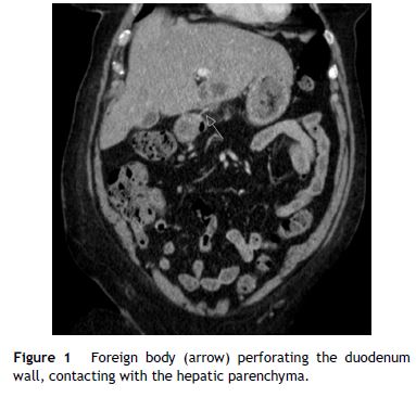
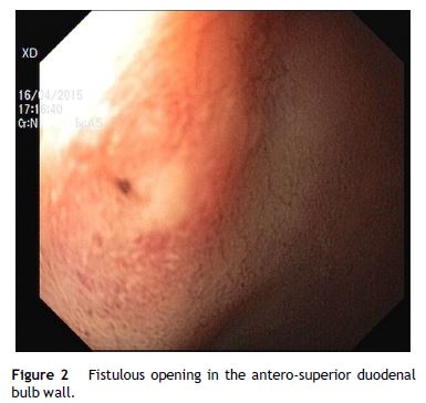
The authors present the case of a 80 years-old woman, with no relevant medical history, which presented to the emergency department for fever (38–39°C), chills, nausea and vomiting, and persistent pain in the right hypochondrium lasting for two days. Initial clinical assessment revealed hypotension, hypoxemia (pO2 < 70 mmHg), slight leukocytosis, CRP 176 mg/L and increased lactates (3.0 mmol). Imaging studies (chest X-ray and ultrasonography) showed no change. The patient was hospitalized in an intermediate care unit where she was started on antibiotics with a diagnosis of sepsis of unknown origin. Two days after, the patient was clinically better, with improvement of the inflammatory parameters, but maintained abdominal complaints and so it was performed an abdominal CT scan where we were able to identify a linear foreign body with approximately 30 mm perforating the intestinal wall at the level of the pylorus, contacting with the hepatic parenchyma where a multiloculated abscess with 44 mm was seen (Fig. 1 (arrow)). After additional conversation with the patient, it was found to be of a fishbone ingested days before. Consequently, an upper endoscopy was performed where it was found a swollen area in the antero-superior duodenal bulb wall with a central fistulous opening (Fig. 2). After discussion with the surgical team it was decided to maintain a conservative strategy (antibiotics plus imaging surveillance). Once there was gradual improvement of the clinical picture, the patient was transferred to the surgery ward and then discharged to outpatient consultation. During the following six months the patient made regular visits, remaining asymptomatic and with imaging resolution of the abscess.
A myriad of ingested sharp-pointed objects have been described. The ones most commonly associated with complications are chicken and fish bones. Patients suspected of swallowing sharp-pointed objects must be evaluated to define the location of the object. However, not always the patients are aware of foreign body ingestion, and in these cases the diagnosis is only made on the occurrence of complications, such as in the reported case. Cases of hepatic abscess due to fish bone penetration are rare and may be fatal.1 Until today, less than 12 cases have been described in the literature. The possible mechanism of the liver abscess secondary to fish bone migration from the duodenum it's the creation of a duodenohepatic fistula covered by duodenal serosa.2 Since the first reported case, treatments usually include drainage of the abscess, removal of the foreign body, and administration of appropriate antibiotics.3 Surviving patients described in previous reports were all surgically treated, except for one case.4 If there is a strong suspicion of bowel perforation by a foreign body or if a foreign body is detected preoperatively, surgery is considered the treatment of choice in current clinical practice, but when the diagnosis is not initially suspected, and if the patient shows a clinically improvement, a conservative approach can be attempted. In our case, once the patient was better under empirical antibiotics it was preferred not to submit the patient to a surgical procedure. Although controversial, the decision turned out to be favorable for the patient as she fully recovered, allowing an outpatient follow-up and six months after discharge she was asymptomatic and the infection fully resolved. So we suggest that medical approaches could be attempted first in such cases. Once the patient experiences signs of clinical deterioration surgical intervention should be promptly considered.
References
1. Chen HK, Kuo JR, Uen YH, Sun DP. Liver abscess secondary to fish bone migration from the duodenum. ANZ J Surg. 2011;81:206. [ Links ]
2. Theodoropoulou A, Roussomoustakaki M, Michalodimitrakis MN, Kanaki C, Kouroumalis EA. Fatal hepatic abscess caused by a fish bone. Lancet. 2002;359:977. [ Links ]
3. Shulda¿s AK, Sumin AV, Tkhorzhevski¿ BB. Pyogenic hepatic abscess developing after perforation of the stomach by a fish bone. Klin Khir. 1992;7:5-6. [ Links ]
4. Yang CY, Kao JH, Liu KL, Chen SJ. Medical treatment of fish bone-related liver abscess. Clin Infect Dis. 2005;41:1689-90. [ Links ]
Ethical disclosures
Protection of human and animal subjects. The authors declare that no experiments were performed on humans or animals for this study.
Confidentiality of data. The authors declare that no patient data appear in this article.
Right to privacy and informed consent. The authors declare that no patient data appear in this article.
Conflicts of interest
The authors have no conflicts of interest to declare.
* Corresponding author.
E-mail address: armandoafp5@gmail.com (A. Peixoto).
Received 12 February 2016; accepted 13 March 2016

