


Serviços Personalizados
Journal
Artigo
 Inglês (pdf)
 Artigo em XML
 Referências do artigo
 Enviar este artigo por email
Indicadores
-
 Citado por SciELO
 -
 Acessos
Links relacionados
-
 Similares em
SciELO
Similares em
SciELO
Compartilhar

 Permalink
GE-Portuguese Journal of Gastroenterology
versão impressa ISSN 2341-4545
GE Port J Gastroenterol vol.22 no.6 Lisboa dez. 2015
https://doi.org/10.1016/j.jpge.2015.07.009
IMAGES IN GASTROENTEROLOGY AND HEPATOLOGY
Duodenal Subepithelial Lesion? An Unexpected Endoscopic Ultrasound Finding
Lesão Subepitelial Duodenal? Um Achado Inesperado na Ecoendoscopia
Susana Marquesa,∗, Pedro Pinto-Marquesb, David Serrab
a Gastroenterology Department, Centro Hospitalar Lisboa Ocidental, Lisbon, Portugal
b Gastroenterology Department, Hospital da Luz, Lisbon, Portugal
* Corresponding author.
Keywords: Duodenal Neoplasms; Endosonography
Palavras-Chave: Neoplasias Duodenais; Ecoendoscopia
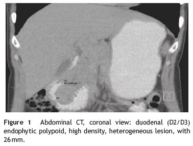
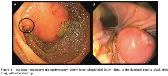
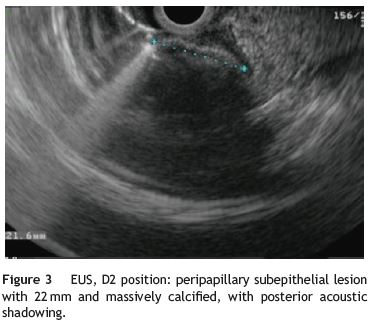
A 39-year-old female with no significant past medical history presented with intense epigastric, colic pain for the three previous days. Laboratory studies showed mild and isolated C-reactive protein (CRP) elevation (1.9 mg/dL); liver function tests and pancreatic enzymes were within normal range. An abdominal computed tomography (CT) was performed and revealed a duodenal (D2/D3) endophytic polypoid, high density, heterogeneous lesion, with 26mm; biliary tree and pancreas were normal (Fig. 1). Upper endoscopy (Fig. 2A) and duodenoscopy (Fig. 2B) identified a 25mm large subepithelial lesion, distal to the duodenal papilla (black circle in Fig. 2A), with ulcerated top, suggestive of gastrointestinal stromal tumor (GIST). Endoscopic ultrasound (EUS) examination confirmed a peripapillary subepithelial lesion with 22mm and massively calcified, with posterior acoustic shadowing (transition zone undefined) (Fig. 3); the common bile duct was normal.
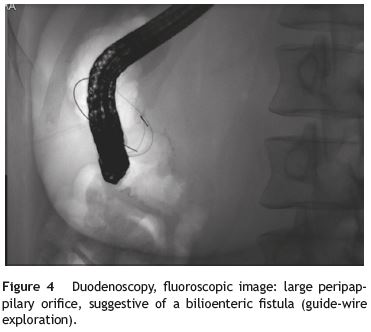
In this patient, EUS findings raised the diagnostic suspicion of a gallstone impacted in the duodenal wall through a peripapillary bilioenteric fistula. Therefore, duodenoscopy was repeated at day 6 but, instead of the lesion previously seen, there was now a large peripappilary orifice, suggestive of a bilioenteric fistula (40mm large after contrast injection and guide-wire exploration) (Fig. 4). There was spontaneous and complete abdominal pain remission in less than a week after admission.
The case presented was particularly challenging as the endoscopic and imagiologic appearance mimicked that of a calcified subepithelial lesion with ulcerated top, such as GIST. Most common subepithelial lesions found in the duodenum are GIST, leyomiomas, lipomas and carcinoids and EUS plays a major role in the differential diagnosis. These lesions may occasionally show calcification, usually focal or punctate.1 However, massive calcification is extremely rare (it has been described in few cases of GIST)1 and may point to an alternative diagnosis such as an impacted gallstone into the duodenal wall, particularly if found in D2. There are only three published case reports of parapapillary fistula with impaction of a gallstone into the duodenal wall.2,3
References
1. Izawa N, Sawada T, Abiko R, Kumon D, Hirakawa M, Kobayashi, et al. Gastrointestinal stromal tumor presenting with prominent calcification. World J Gastroenterol. 2012;18:5645-8. [ Links ]
2. Okabe Y, Kaji R, Ishida Y, Noda T, Sasaki Y, Tsuruta O, et al. Successful endoscopic extraction of a large impacted choledocholithiasis in the ampulla of vater: two interesting cases. Dig Endosc. 2010;22:103-6. [ Links ]
3. Papanikolaou IS, Polymeros D, Kontopoulou C, Chatzilia D, Triantafyllou K. Impaction of a gallstone into the duodenal wall. Endoscopy. 2010;42:297-8. [ Links ]
* Corresponding author.
E-mail address: xsusanamarx@gmail.com (S. Marques).
Conflicts of interest
The authors have no conflicts of interest to declare.
Disclosures
All authors have approved the manuscript and agree with its submission to GE - Portuguese Journal of Gastroenterology. All authors have nothing to disclose.
Author contributions
Susana Marques did the writing of the manuscript. Pedro Pinto-Marques and David Serra were responsible for the revision of its contents.
Received 16 June 2015; accepted 6 July 2015

