


Serviços Personalizados
Journal
Artigo
 Inglês (pdf)
 Artigo em XML
 Referências do artigo
 Enviar este artigo por email
Indicadores
-
 Citado por SciELO
 -
 Acessos
Links relacionados
-
 Similares em
SciELO
Similares em
SciELO
Compartilhar

 Permalink
GE-Portuguese Journal of Gastroenterology
versão impressa ISSN 2341-4545
GE Port J Gastroenterol vol.22 no.5 Lisboa out. 2015
https://doi.org/10.1016/j.jpge.2015.05.002
IMAGES IN GASTROENTEROLOGY AND HEPATOLOGY
Endoscopic Ultrasound and Anal Pain: The Key to Diagnosis
Ecoendoscopia e Dor Anal: A Chave do Diagnóstico
Tarcísio Araújoa,∗, Fernando Castro-Poçasa,b,c, Isabel Pedrotoa,c
a Gastroenterology Department, Centro Hospitalar do Porto, Porto, Portugal
b Ultrasound Department, Centro Hospitalar do Porto, Porto, Portugal
c Institute of Biomedical Sciences Abel Salazar, University of Porto, Porto, Portugal
* Corresponding author.
Keywords: Anus Diseases; Endosonography; Pain
Palavras-Chave: Doenças do Ânus; Dor; Ecoendoscopia
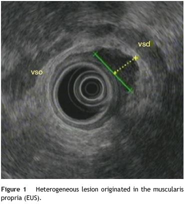
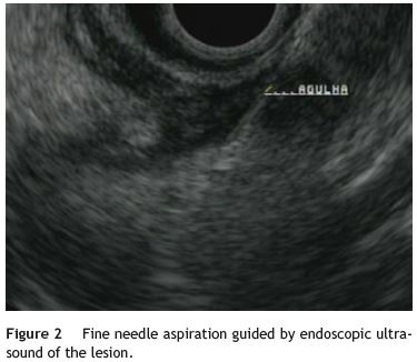
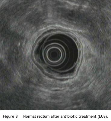
A 73-year-old male patient with a history of pulmonary tuberculosis in childhood, a gastric adenocarcinoma (intestinal type) T1N0M0 (TNM classification), submitted to a partial gastrectomy Roux-en-Y, 10 years before, and a T1 lowgrade papillary bladder urothelial carcinoma treated with a transurethral resection and epirubicin, 1 year before, reported anal pain and fever for the last 5 days. The patient denied diarrhea, hematochezia or weight loss. In the anoscopy a painful bulging lesion with 2-3 cm was detected in distal rectum. Anorectal endoscopic ultrasonography (EUS) showed a heterogeneous, irregular lesion in the distal right rectal wall, with a central hypoechogenic area measuring 26mm on its longest axis and 14mm on its shortest axis, originated in the muscularis propria (Fig. 1). We performed a (ultrasound guided) fine needle (22 gauge) aspiration (FNA), three passages, with collection of solid and fluid material (Fig. 2). The patient did prophylaxis with ciprofloxacin for 5 days after the procedure. Culture of fluid revealed polymicrobial flora, probably contamination. Cytology revealed the presence of leukocytes and necrosis material, supporting the hypothesis of rectal abscess. The drainage of the abscess was proposed to the patient, but he refused the procedure. An antibiotic treatment with trimethoprim-sulfamethoxazole was given during 14 days, with resolution of symptoms. After 1 month, another endoscopic ultrasonography showed no lesions (Fig. 3).
1. Commentary
Perirectal abscesses are frequently the manifestation of an infected anal gland,1 usually originating anal pain.2 In this case, anorectal abscess was the most probable diagnosis, so anorectal EUS was performed. Nevertheless the image was atypical and the internal orifice or fistula was not observed, so FNA was executed to achieve a definitive diagnosis, which was supported by the cytology sample. Drainage is the first choice for therapy,3 but in this case, because the patient refused that solution, we chose to treat him only with antibiotics, achieving an effective management of the abscess.
We emphasize the image of this perirectal abscess and the importance of fine needle aspiration for the diagnosis of atypical rectal abscess.
References
1. Whiteford M. Perianal abscess/fistula disease. Clin Colon Rectal Surg. 2007;20:102-9. [ Links ]
2. Marcus R, Stine R, Cohen M. Perirectal abscess. Ann Emerg Med. 1995;25:597-603. [ Links ]
3. Whiteford M, Kilkenny J III, Hyman N, Buie W, Cohen J, Orsay C, et al. Practice parameters for the treatment of perianal abscess and fistula-in-ano. Dis Colon Rectum. 2005;48:1337-42. [ Links ]
* Corresponding author.
E-mail address: araujo.tarcisio@gmail.com (T. Araújo).
Ethical disclosures
Protection of human and animal subjects. The authors declare that no experiments were performed on humans or animals for this study.
Confidentiality of data. The authors declare that they have followed the protocols of their work center on the publication of patient data.
Right to privacy and informed consent. The authors declare that no patient data appear in this article.
Conflicts of interest
No conflicts of interest to declare.
Received 11 February 2015; accepted 1 May 2015

