


Serviços Personalizados
Journal
Artigo
 Inglês (pdf)
 Artigo em XML
 Referências do artigo
 Enviar este artigo por email
Indicadores
-
 Citado por SciELO
 -
 Acessos
Links relacionados
-
 Similares em
SciELO
Similares em
SciELO
Compartilhar

 Permalink
GE-Portuguese Journal of Gastroenterology
versão impressa ISSN 2341-4545
GE Port J Gastroenterol vol.22 no.4 Lisboa ago. 2015
https://doi.org/10.1016/j.jpge.2015.04.006
IMAGES IN GASTROENTEROLOGY AND HEPATOLOGY
Duodenal and Colonic Metastases of Ovarian Neoplasm
Metástases Duodenais e Cólicas de Neoplasia do Ovário
Sandra Barbeiro∗, Catarina Atalaia Martins, Cláudia Gonçalves
Gastroenterology Department, Centro Hospitalar de Leiria, Leiria, Portugal
* Corresponding author.
Keywords: Colonic Neoplasms/secondary; Duodenal Neoplasms/secondary; Ovarian Neoplasms
Palavras-chave: Neoplasias do Cólon/secundária; Neoplasias do Duodeno/secundária; Neoplasias do Ovário
1. Case report
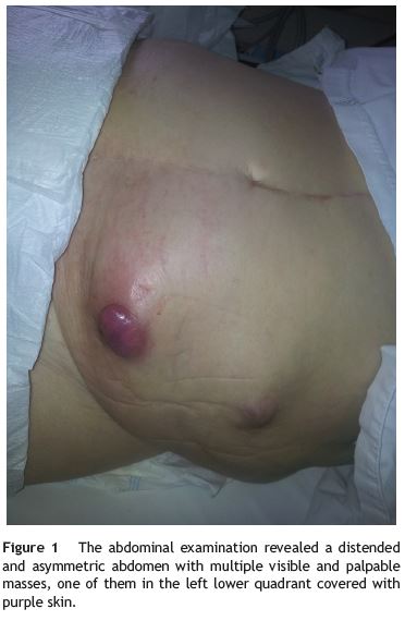
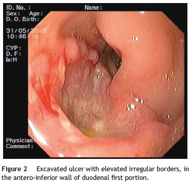
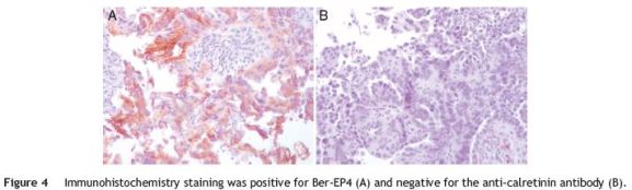
A 75-year-old female presented with asthenia and melena. Her past medical history included Alzheimers disease, laparotomy in childhood due to unknown reason and an advanced ovarian neoplasm with peritoneal carcinomatosis in palliative treatment during the last year. Two years before, the patient was submitted to gastrointestinal endoscopic examination (endoscopy and colonoscopy) that were normal. She was pale and her vital signs were stable. The abdomen was distended and asymmetric, with an abdominal surgical scar in midline and multiple visible and palpable masses in all quadrants (Fig. 1). Laboratory tests showed hemoglobin of 8.3 g/dL. Endoscopy demonstrated an excavated ulcer with elevated irregular borders, in the antero-inferior wall of duodenal first portion with no major bleeding stigmata (Fig. 2) and endoscopic biopsies were performed. Blood transfusion and medical treatment were started and clinical improvement was observed. However, on the third day after admission she had haematochezia. A colonoscopy was performed and showed two ulcerated masses in the sigmoid colon with active bleeding (Fig. 3). Endoscopic hemostasis with argon plasma coagulation was attempted without success. Biopsies were done. Histopathological examination confirmed metastatic malignant ovarian neoplasm in duodenum and colon (Fig. 4).

2. Discussion
Duodenal and colonic metastases are a very rare cause of gastrointestinal bleeding.1,2 However, they should be considered in a patient presenting with gastrointestinal bleeding and a previous history of malignancy.1 The main dissemination pattern in epithelial ovarian neoplasm is the transcoelomic route. Metastasis to the intestinal mucosa can occur as a result of transmural involvement by invasion from the serosal surface. Lymphatic and hematogenous dissemination may also occur in the case of advanced peritoneal disease.1 Symptoms are nonspecific and similar to those caused by other gastrointestinal tumors: abdominal pain, altered bowel habits, tenesmus, small bowel obstruction or perforation, hematemesis, melena and anemia. Imaging and endoscopic findings are unspecific and histopathological examination allowed definitive diagnosis. Special immunohistochemical staining, including cytokeratin-7 and 20, Ber-EP4 and the anti-calretinin antibody, is important to confirm diagnosis.1,3-5 Prognosis is poor and treatment is mainly palliative. Endoscopic treatment is hardly effective. In some cases surgical resection and chemotherapy may be considered.2
References
1. Loualidi A, Spooren PF, Grubben MJ, Blomjous CE, Goey SH. Duodenal metastasis: an uncommon cause of occult small intestinal bleeding. Neth J Med. 2004;60:201-5. [ Links ]
2. Zighelboim I, Broaddus R, Ramirez P. Atypical sigmoid metastasis from a high-grade mixed adenocarcinoma of the ovary. Gynecol Oncol. 2004;94:850-3. [ Links ]
3. Okamoto S, Ito K, Sasano H, Moriya T, Niikura H, Terada Y, et al. Ber-EP4 and anti-calretinin antibodies: a useful combination for differential diagnosis of various histological types of ovarian cancer cells and mesothelial cells. Tohoku J Exp Med. 2005;206:31-40. [ Links ]
4. Shin JH, Bae JH, Lee A, Jung CK, Yim HW, Park JS, et al. CK7, CK20, CDX2 and MUC2 immunohistochemical staining used to distinguish metastatic colorectal carcinoma involving ovary from primary ovarian mucinous adenocarcinoma. Jpn J Clin Oncol. 2010;40:208-13. [ Links ]
5. Tot T. Cytokeratins 20 and 7 as biomarkers: usefulness in discriminating primary from metastatic adenocarcinoma. Eur J Cancer. 2002;38:758-63. [ Links ]
* Corresponding author.
E-mail address: sandrabarbeiro@gmail.com (S. Barbeiro).
Ethical disclosures
Protection of human and animal subjects. The authors declare that no experiments were performed on humans or animals for this study.
Confidentiality of data. The authors declare that no patient data appear in this article.
Right to privacy and informed consent. The authors declare that no patient data appear in this article.
Conflicts of interest
The authors have no conflicts of interest to declare.
Received 28 February 2015; accepted 25 April 2015

