


Serviços Personalizados
Journal
Artigo
 Inglês (pdf)
 Artigo em XML
 Referências do artigo
 Enviar este artigo por email
Indicadores
-
 Citado por SciELO
 -
 Acessos
Links relacionados
-
 Similares em
SciELO
Similares em
SciELO
Compartilhar

 Permalink
GE-Portuguese Journal of Gastroenterology
versão impressa ISSN 2341-4545
GE Port J Gastroenterol vol.22 no.3 Lisboa jun. 2015
https://doi.org/10.1016/j.jpge.2015.01.005
IMAGES IN GASTROENTEROLOGY AND HEPATOLOGY
Rapid Progression of Multifocal Hepatic Epithelioid Hemangioendothelioma
Rápida Progressão de um Hemangioendotelioma Epitelióide Hepático Multifocal
João Ressurreiçãoa,b,∗, Celso Matosb, Maria Balib
a Radiology Department, Centro Hospitalar de Vila Nova de Gaia/Espinho, Vila Nova de Gaia, Portugal
b Radiology Department, Hôpital Erasme, Université Libre de Bruxelles, Brussels, Belgium
* Corresponding author.
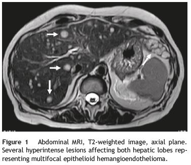
A 60-year-old woman with past medical history of chronic pancreatitis and bilioenteric anastomosis was referred to our institution for routine unenhanced pancreatic MR evaluation. Besides the expected findings of uncomplicated chronic pancreatitis, the study revealed several focal lesions distributed through the liver, suggestive of metastases. They were hypointense in T1 imaging and remarkably hyperintense in T2-weighted images (Fig. 1). Blood tests where mostly unexceptional, only revealing elevated gammaglutamyl transferase (88 U/L (normal: 5-36 U/L)).
A liver biopsy was performed and, despite the obtention of only a small amount of diagnostic material, it was still possible to exclude carcinomatous, neuroendocrine and hepatocytary origin and to detect positivity for CD34, a marker present in tumors of hematopoietic or endothelial lineage.1
PET-CT was also realized for staging, demonstrating multiple slightly hypermetabolic liver lesions and significant hypermetabolism centered in the left aryepiglottic fold. After biopsy the neck lesion was diagnosed as squamous-cell carcinoma, non-related to the liver lesions. Neck MRI staging revealed invasion of the ipsilateral piriform sinus and the presence of an ipsilateral level IV necrotic lymph node (TNM: T2,N1,M0).
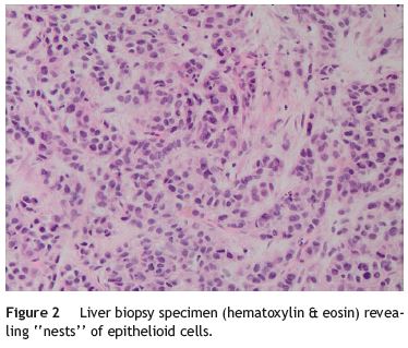
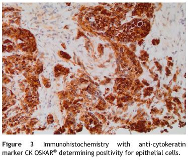
With these results the possibility of a primary, multifocal, vascular liver malignancy started to gain strength and another liver biopsy was taken. The samples were representative and histological analysis revealed the presence of nests of epithelioid cells (Fig. 2); the immunohistochemical tests detected positivity for anti-cytokeratin marker (CK OSKAR®), again suggesting the epithelial nature of the lesion (Fig. 3). Altogether, the findings of both biopsies were highly suggestive of hepatic epithelioid hemangioendothelioma (EHE).
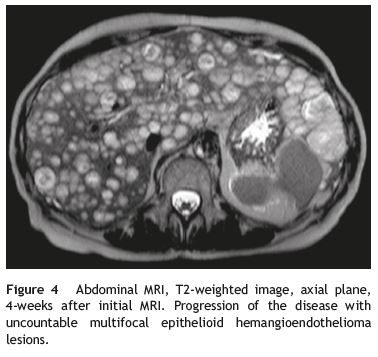
A final contrast-enhanced abdominal MRI (Fig. 4) was taken just before treatment was initialized (4 weeks after the first MRI). The liver parenchyma was now almost completely substituted by hemangioendothelioma lesions that reveal progressive contrast enhancement in the portal and delayed imaging phases.
Due to massive hepatic involvement, liver transplantation would be the best therapeutic option.2 However, the synchronous laryngeal tumor precluded this alternative and the patient started a regime of IV bevacizumab, an angiogenesis inhibitor. After 2 months of clinical and tumoral stability (MRI proven), patients clinical condition worsened and a new MRI revealed progression of liver disease and ascites. The patient finally died from respiratory infectious complication.
HEH is a very uncommon primary tumor of liver vasculature, usually defined as intermediate-grade malignancy and considered one of the less aggressive hepatic malignancies. Nevertheless, there are aggressive cases with progressive tumor growth leading to hepatic failure, extra-hepatic metastases and death.3
Three forms of the tumor are recognized: single nodular, multifocal and diffuse type. The majority of tumors correspond to the multifocal-type. Characteristic MR features of multifocal HEH include multiple hepatic nodules usually centered in the periphery of the liver, coalescent, hypointense in T1-weighted imaging, heterogeneously hyperintense in T2-weighted imaging with progressive, delayed contrastenhancement.3
When feasible, the most effective treatment is liver transplantation with 1-year and 5-year patient survival rates of 96% and 54.5%.4 Evidence suggests that even patients with limited extra-hepatic disease can be considered for transplantation. Other therapeutic strategies, as the use of angiogenesis inhibitors, can be considered when transplantation is not an option.5
References
1. Traweek ST, Kandalaft PL, Mehta P, Battifora H. The human hematopoietic progenitor cell antigen (CD34) in vascular neoplasia. Am J Clin Pathol. 1991;96:25-31. [ Links ]
2. Madariaga JR, Marino IR, Karavias DD, Nalesnik MA, Doyle HR, Iwatsuki S, et al. Long-term results after liver transplantation for primary hepatic hemangioendothelioma. Ann Surg Oncol. 1995;2:483-7. [ Links ]
3. Lyburn ID, Torreggiani C, Harris AC, Zwirewich CV, Buckley AR, Davis JE, et al. Epithelioid hemangioendothelioma: sonographic CT, and MR imaging appearances. AJR Am J Roentgenol. 2003;180:1359-64. [ Links ]
4. Mehrabi A, Kashfi A, Fonouni H, Schemmer P, Schmied BM, Hallscheidt P, et al. Primary malignant hepatic epithelioid hemangioendothelioma: a comprehensive review of the literature with emphasis on the surgical therapy. Cancer. 2006;107:2108-211. [ Links ]
5. Emamaullee JA, Edgar R, Toso C, Thiesen A, Bain V, Bigam D, et al. Vascular endothelial growth factor expression in hepatic epithelioid hemangioendothelioma: Implications for treatment and surgical management. Liver Transpl. 2010;16:191-7. [ Links ]
* Corresponding author.
E-mail address: joaofres@gmail.com (J. Ressurreição).
Ethical disclosures
Protection of human and animal subjects. The authors declare that no experiments were performed on humans or animals for this study
Confidentiality of data. The authors declare that no patient data appear in this article
Right to privacy and informed consent. The authors declare that no patient data appear in this article.
Conflicts of interest
The authors have no conflicts of interest to declare.
Received 6 December 2014; accepted 5 January 2015
Acknowledgements
The authors are most grateful to Professor Pieter Demetter (Department of Pathology, Hôpital Erasme, Brussels, Belgium) for his important contribution to the accomplishment of this work. The pathological findings were crucial for the final diagnosis, particularly due to the lack of specificity of the imaging findings. The diagnostic and therapeutic decisions were taken together, in group, by different members of the Departments of Gastroenterology and Hepato-Pancreatology, Medical Oncology and Digestive Surgery of the Hôpital Erasme, to whom we also would like to thank.

