


Serviços Personalizados
Journal
Artigo
 Inglês (pdf)
 Artigo em XML
 Referências do artigo
 Enviar este artigo por email
Indicadores
-
 Citado por SciELO
 -
 Acessos
Links relacionados
-
 Similares em
SciELO
Similares em
SciELO
Compartilhar

 Permalink
GE-Portuguese Journal of Gastroenterology
versão impressa ISSN 2341-4545
GE Port J Gastroenterol vol.22 no.1 Lisboa fev. 2015
https://doi.org/10.1016/j.jpge.2014.10.003
CLINICAL CASE
Accidental Ingestion of Dentistry Material - Report of Cases and Challenges from the Pediatrician Point of View
Ingestão Acidental de Material Dentário - Casos Clínicos e Desafios do Ponto de Vista do Pediatra
Joana Cotrima,∗, Susana Corujeirab, Joana Jardimb, Hélder Cardosoc, Eunice Trindaded, Jorge Amil Diasd
a Paediatrics Department, Hospital de Vila Real, Centro Hospitalar de Trás-os-Montes e Alto Douro E.P.E., Vila Real, Portugal
b Paediatrics Department, Hospital de São João, Centro Hospitalar de São João E.P.E., Porto, Portugal
c Gastroenterology Department, Hospital de São João, Centro Hospitalar de São João E.P.E., Porto, Portugal
d Paediatric Gastroenterology Unit, Paediatrics Department, Hospital de São João, Centro Hospitalar de São João E.P.E., Porto, Portugal
* Corresponding author.
ABSTRACT
Introduction: Aspiration or ingestion of foreign bodies may occur during dental procedures. Diagnosis and management of these accidents is sometimes challenging. The authors present a small series of clinical cases:
Case 1: Adolescent observed due to suspected accidental bracket ingestion, not visible on x-ray, removed by upper digestive endoscopy.
Case 2: Adolescent observed after accidental ingestion of a dental file. Conflicting results in image exams and absence of object progression led to enteroscopy for extraction.
Case 3: Adolescent observed due to accidental ingestion of a surgical blade, visualized on image study but not accessible by endoscopy, resulting in latter spontaneous elimination.
Discussion: Image study is frequently useful when metallic object ingestion is suspected, but has some limitations. In some cases, mucosal protections must be used during removal procedures. Prevention of such accidents is the best approach, using appropriate protections to secure airway and digestive tract during dental procedures.
Keywords: Dental Care; Endoscopy; Foreign Bodies; Gastrointestinal Tract
RESUMO
Introdução: A aspiração ou ingestão de corpos estranhos pode ocorrer durante qualquer procedimento dentário. O diagnóstico e a resolução destas situações podem colocar desafios. Os autores apresentam três casos clínicos:
Caso 1: Adolescente observada por suspeita de ingestão acidental de um bracket de aparelho fixo de ortodontia, não visualizado na radiografia e removido por endoscopia digestiva alta.
Caso 2: Adolescente com história de ingestão acidental de uma lima de dentista. Os exames complementares realizados não foram esclarecedores e, dado não haver progressão do corpo estranho, foi necessária a sua remoção por enteroscopia.
Caso 3: Adolescente que recorre à urgência por ingestão acidental de uma lâmina de dentista, inacessível por endoscopia e que acabou por ser eliminada espontaneamente.
Discussão: Os exames imagiológicos são habitualmente úteis na suspeita de ingestão de corpos estranhos metálicos mas a informação que fornecem é limitada. A natureza cortante do material dentário deglutido exige que se tomem cuidados adicionais na sua remoção, utilizando auxiliares protectores. A prevenção destes acidentes deve ser promovida, recorrendo a protecções da via aérea e digestiva durante a realização de procedimentos dentários.
Palavras-Chave: Corpos Estranhos; Cuidados Dentários; Endoscopia; Tracto Gastrointestinal
1. Introduction
Ingestion of foreign bodies may occur during dental procedures.1-4 Orthodontic components are mostly small, handling can be difficult and any object that is placed into or removed from the oral cavity can be aspirated or ingested.2,4 Supine or semi-recumbent position of the patient can increase the risk for such accidents; moreover, oral cavity is usually of limited access and visibility.2 Diagnosis and management of foreign objects ingestion is sometimes challenging. Depending on the features of ingested material, these accidents can present minimal danger or need urgent medical evaluation.2-5
After accidental ingestion, 75% of foreign bodies pass the gastrointestinal tract spontaneously.6 However, it is known that sharp and pointed objects are associated with higher complication rates, such as perforation of the gastrointestinal tract, hemorrhage and ulceration. Depending on anatomic location and the objects features, urgent endoscopic evaluation may be necessary. The aim of this article is to present three cases of accidental ingestion of dentistry materials in previously healthy patients, to draw attention to the difficulties in diagnosing and managing these accidents and to the need to prevent them.
2. Cases report
2.1. Case 1
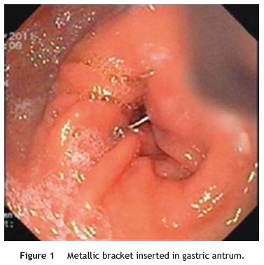
A seventeen-year-old adolescent girl was observed in the emergency department after suspected accidental ingestion of a bracket from her dental braces, during sleep. The only symptom was abdominal pain. Initial evaluation included chest and abdominal X-rays, but no foreign object was visualized and there were no radiological signs of intestinal perforation or occlusion. Due to the persistence of symptoms, upper digestive endoscopy (UDE) was performed and it revealed a metallic object compatible with the braces bracket inserted in the gastric antrum mucosa (Fig. 1). The metallic bracket, 3mm×4mm, was successfully removed using an endoscopy retrieval net and a small ulceration was identified in the gastric mucosa.
2.2. Case 2
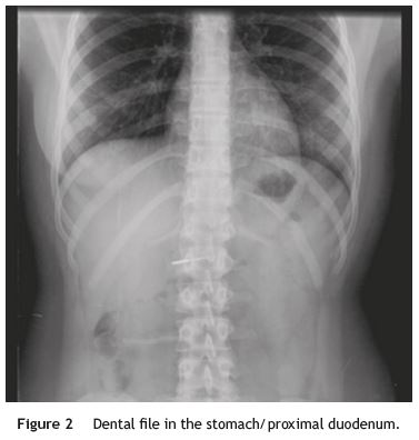
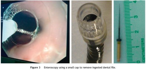
A previously healthy 13-year-old adolescent girl was admitted to the emergency department after accidental ingestion of a dental file during a dental procedure. She had no symptoms. The X-rays revealed a metallic object apparently located on the stomach/duodenal arch (Fig. 2). It was not accessible on the UDE. The patient was discharged from the emergency department and revaluated in the ambulatory clinic. During follow-up, she remained asymptomatic and spontaneous elimination of the dental file was not observed. Control radiographs suggested no progression of object. Abdominal CT scan was used to determine the exact location of the dental file, suggesting that it was entrapped in the distal duodenum/proximal jejunum. Balloon enteroscopy was needed to extract the foreign body, which was found inserted in the mucosa of the second part of the duodenum. A 4 cm long dental file was extracted to the intestinal lumen and carefully to the exterior, using a small cap in order to prevent mucosal damage during the removal procedure (Fig. 3). A small ulceration in the duodenal mucosa was visualized after the object was removed.
2.3. Case 3
A fifteen-year-old male was observed in the emergency department after accidental ingestion of a surgical blade (1.5 cm long) during a dental procedure. He presented no symptoms. The abdominal X-ray suggested location of the foreign object on the proximal small intestine, but no object was visualized on UDE, which included visualization of the second portion of the duodenum. Considering the dangerous nature of the blade, pointed and sharp, medical revaluation was warranted. During follow-up, the patient remained asymptomatic but spontaneous elimination was not observed. Image study was repeated (posteroanterior and lateral X-rays), showing object progression through the intestinal tract and, by the fourth day after accidental ingestion, no foreign object was identified on X-ray. During the posterior revaluations in the ambulatory clinic the patient remained asymptomatic and it was considered that the foreign object had been eliminated.
3. Discussion
Doctors and patients must be aware that ingestion or aspiration of dentistry material is a potential risk in dental procedures. The reported incidence of ingested objects of dental origin varies considerably in literature, between 3.6% and 27.7% of all foreign bodies.7
Any object that is routinely removed from or placed into the oral cavity should be carefully manipulated. All orthodontic components must be adequately fixed and adapted, preventing accidental aspiration or ingestion.2 There are many strategies to avoid such accidents during dental procedures: use of rubber dam, use of gauze throat, tieing small objects with floss, directly observing the entire procedure, using the most upright patient position possible and providing detailed instructions to patients.2,3
When the accident occurs, health professionals must be aware of a correct protocol to manage these situations, in order to provide timely and adequate intervention.2-4
Image studies are the initial approach and are very useful in determining the location of ingested foreign bodies. Plain film radiograph is the first diagnostic study, however, it has some limitations: radiolucent material is not visible on Xray, small metallic objects may not be identified and object location may be inaccurate.2,5
After ingestion, 75% of foreign bodies pass the gastrointestinal tract spontaneously.6 However, management varies based upon the features of the object ingested and its location. Esophageal foreign bodies may result in complete esophageal obstruction or perforation with life-threatening risks, and therefore, they should always be removed. When objects reach the stomach, the majority will pass the gastrointestinal tract without complications, as a result of peristaltic movement. However, long standing foreign bodies or those that are sharp and pointed may become impacted in the gastric mucosa and result in inflammation, ulceration, hemorrhage or perforation. Foreign objects that progress to the small bowel typically cause no symptoms and spontaneous elimination time varies from 7 to 10 days. If the object fails to progress, device-assisted enteroscopy techniques or surgical intervention should be considered.
All cases described in this series could have been prevented and, especially cases 2 and 3, imply major potential risks due to the features of ingested objects.
Presented cases emphasize the need for an individual approach in each case of accidental foreign object ingestion and the challenge of an accurate diagnosis.
Case 1 draws attention to the role of upper digestive endoscopy in symptomatic patients even with normal X-rays.
Regarding treatment, UDE is the main option in case of foreign body ingestion. However, the object may not be within the reach of the endoscope and other techniques may be required. Enteroscopy allows access to a larger extent of intestinal tract and it should be considered as a therapeutic option. Several devices may be used to facilitate endoscopic management of foreign objects, including overtubes, caps and hoods to protect the gastrointestinal mucosa during removal of sharp objects, as occurred in case 2.6
Case 3 reveals the importance of serial radiograph studies in select patients. In case of ingestion of sharp objects, perforation rates are as high as 35%.6 If the patient is asymptomatic and the object has passed beyond the duodenum, daily control radiographs should be obtained to monitor object progression and search for signs of complications, such as the presence of free air. In case of suspected complications or absence of progression through the digestive tract surgical management may be warranted. The authors present three different cases of accidental ingestion in dental practice and three different approaches on the diagnosis and management of these accidents. Overall, prevention of accidental ingestion of dentistry material is still the best approach.
References
1. Bhatnagar S, Das U, Chandan G, Prashanth S, Gowda L, Shiggaon N. Foreign body ingestion in dental practice. J Indian Soc Pedod Prev Dent. 2011;29:336-8. [ Links ]
2. Milton T, Hearing S, Ireland A. Ingested foreign bodies associated with orthodontic treatment: report of three cases and review of ingestion/aspiration management. Br Dent J. 2001;190:592-6. [ Links ]
3. Obinata K, Satoh T, Towfik A, Nakamura M. An investigation of accidental ingestion during dental procedures. J Oral Sci. 2011;53:495-500. [ Links ]
4. Parolia A, Kamath M, Kundubala M, Manuel T, Mohan M. Management of foreign body aspiration or ingestion in dentistry. Kathmandu Univ Med J. 2009;7:165-71. [ Links ]
5. Michaud L, Bellaiche M, Olives J. Groupe Francophone d´hépatologie, gastroenterology et nutrition pédiatriques (GFHGNP). Ingestion of foreign bodies in children. Recommendations of the French - Speaking Group of Pediatric Hepatology, Gastroenterology and Nutrition. Arch Pediatr. 2009;16(1):54-61. [ Links ]
6. Christopher C, Thompson MD. Gastrointestinal foreign bodies. In: Current, diagnosis and treatment - gastroenterology, hepatology, and endoscopy. 1st ed New York: McGraw-Hill Companies; 2009. p. 371-4. [ Links ]
7. Umesan U, Chua K, Balakrishnan P. Prevention and management of accidental foreign body ingestion and aspiration in orthodontic practice. Ther Clin Risk Manag. 2012;8:245-52. [ Links ]
*Corresponding author
E-mail address: joana_cotrim@hotmail.com (J. Cotrim).
Ethical disclosures
Protection of human and animal subjects. The authors declare that no experiments were performed on humans or animals for this study.
Confidentiality of data. The authors declare that they have followed the protocols of their work center on the publication of patient data
Confidentiality of data. The authors declare that no patient data appear in this article.
Received 2 June 2014; accepted 15 October 2014

