


Serviços Personalizados
Journal
Artigo
 Inglês (pdf)
 Artigo em XML
 Referências do artigo
 Enviar este artigo por email
Indicadores
-
 Citado por SciELO
 -
 Acessos
Links relacionados
-
 Similares em
SciELO
Similares em
SciELO
Compartilhar

 Permalink
Acta Obstétrica e Ginecológica Portuguesa
versão impressa ISSN 1646-5830
Acta Obstet Ginecol Port vol.12 no.3 Coimbra set. 2018
ISSUE IMAGE/IMAGEM DO TRIMESTRE
Fetal intra-abdominal umbilical vein varix
Dilatação da veia umbilical fetal
Ana Isabel Correia*, Fátima Leitão**, Sara Neto***
Centro Hospitalar do Baixo Vouga – Departamento da Saúde da Mulher e da Criança.
*Interna Formação Específica
**Assistente Hospitalar Graduada
***Assistente Hospitalar Graduada Sénior
Endereço para correspondência | Dirección para correspondencia | Correspondence
ABSTRACT
Fetal umbilical intra-abdominal vein varix is a rare congenital malformation characterized by focal dilatation of the umbilical vein. It's clinical significance is unclear and is usually an isolated finding, in some cases it may be associated with other fetal anomalies, chromosomal defects or fetal death. The authors report a case of pregnant woman at 25 weeks of gestation with a fetus affected by dilatation of an intraabdominal portion of the umbilical vein, with a favorable progress and outcome.
Keywords: Antenatal ultrasound; Fetal anomalies; Umbilical vein varix.
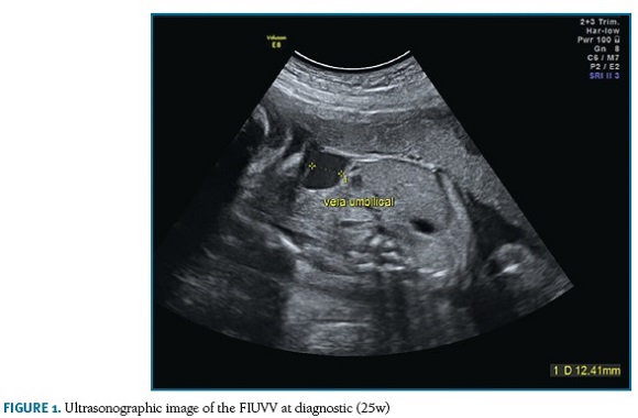
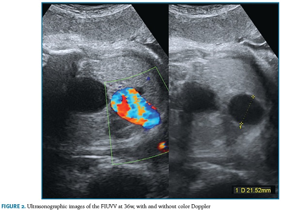
Fetal umbilical intra-abdominal vein varix is a rare congenital malformation characterized by focal dilatation of the umbilical vein. The diagnosis is made by prenatal sonographic diagnosis of a umbilical vein diameter superior to the parameters established for a given gestational age. Although it's clinical significance is unclear and is usually an isolated finding, in some cases it may be associated with other fetal anomalies, chromosomal defects or fetal death. Most cases progress favourably, however, thrombosis is a possible complication that can result in an adverse outcome. The authors report a case of pregnant woman at 25 weeks of gestation with a fetus affected by dilatation of an intra-abdominal portion of the umbilical vein. The combined screening for fetal aneuploidy performed at 12 weeks of gestation reported a low risk, the morphological ultrasound showed a female fetus with a normal anatomical study. It was found an extrahepatic focal dilation of the intra-abdominal portion of the umbilical vein with 12 mm in diameter at 25 weeks (Figure 1), which reached a maximum diameter of 21 mm at 36 weeks (Figure 2). The serial controls showed evolution without impact on fetal hemodynamic status. Labor was induced at 38 weeks of gestation, giving birth, by instrumental delivery, a 3740g female baby. No abnormalities were evident in the physical examination. Abdominal ultrasound performed on the newborn were normal.
BIBLIOGRAPHY
1. Virdis G et al. Umbilical intra-abdominal vein varix: a case report and review of the literature. Clin Exp Obstet Gynecol. 2016;43(2):268-270. [ Links ]
2. Beraud E, Rozel C, Milon J, Darnault P. Umbilical vein varix: Importance of ante- and post-natal monitoring by ultrasound. Diagn Interv Imaging. 2015;96(1):21-26. [ Links ]
3. Sanapo L, Burul G, Saccardi C, Nardelli GB, D'Antona D. Four cases of fetal intra-abdominal umbilical vein varix: a single centre's approach to management. J Obstet Gynaecol 2013;33(4):375-377. [ Links ]
4. Mankuta D, Nadjari, M, Pomp G. Isolated Fetal Intra-Abdominal Umbilical Vein Varix - Clinical Importance and Recommendations. J Ultrasound Med 2011;30:273–276. [ Links ]
5. Navarro-González T et al. Perinatal outcome after prenatal diagnosis of intra-abdominal umbilical vein varix. Ginecol Obstet Mex 2013;81(3):140-145. [ Links ]
Endereço para correspondência | Dirección para correspondencia | Correspondence
Ana Isabel Correia
Centro Hospitalar Baixo Vouga
Aveiro, Portugal
E-Mail: anacorreia87@gmail.com
Recebido em: 25/06/2017
Aceite para publicação: 18/09/2017

