Services on Demand
Journal
Article
Indicators
-
 Cited by SciELO
 -
 Access statistics
Related links
-
 Similars in
SciELO
Share
Nascer e Crescer
Print version ISSN 0872-0754On-line version ISSN 2183-9417
Nascer e Crescer vol.28 no.2 Porto June 2019
https://doi.org/10.25753/BirthGrowthMJ.v28.i2.15862
WHAT IS YOUR DIAGNOSIS? | QUAL O SEU DIAGNÓSTICO?
Stomatological clinical case
Caso clínico estomatológico
Sofia PiresI, Flávia BelinhaI
I Hospital Pediátrico de Coimbra, Centro Hospitalar e Universitário de Coimbra. 3000-602 Coimbra, Portugal. sofia.pires88@gmail.com; flaviaoliveirabelinha@gmail.com
Endereço para correspondência | Dirección para correspondencia | Correspondence
ABSTRACT
The case of a newborn with a gingival mass corresponding to a natal tooth is reported. In this rare and idiopathic disorder, teeth are present at birth. When they show increased mobility and/or complications are present (feeding difficulties, laceration of the mother`s nipples), teeth should be extracted.
Keywords: gingival mass; natal tooth; newborn
RESUMO
Os autores descrevem o caso de um recém-nascido com uma massa gengival correspondente a um dente natal. Neste distúrbio raro e idiopático, os dentes estão presentes ao nascimento. Quando têm uma grande mobilidade e/ou complicações associadas (dificuldades na alimentação, laceração dos mamilos da mãe), devem ser extraídos.
Palavras-chave: massa gengival; dente natal; recém-nascido
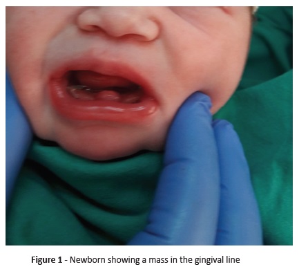
A female infant was born at 38 weeks of gestation. On physical examination at birth, a mass in the midline maxillary gum line was noticed (Figure 1). The mass was mobile to touch and firmly consistent. Physical examination was otherwise unremarkable.
During first postnatal days, the newborn evidenced no breastfeeding problems. She was discharged from the hospital at day three of life and referenced to a Stomatology consultation. At the age of five days the tooth became visible, being removed in the Stomatology clinic with no bleeding problems.
What is the diagnosis?
Diagnoses
Natal tooth
Discussion
Natal and neonatal teeth are rare disorders of tooth eruption, with incidence varying from 1:2.000 to 1:3.000.1 Natal teeth are present at birth and neonatal teeth erupt within the first month of life. The former are more frequent (3 versus 1), and both are more frequent in females.1,2 Althoughtheseconditions’ exact etiology is unknown, they have been associated with various syndromes, including chondroectodermal dysplasia (Ellis Van Creveld syndrome), Pierre Robin syndrome, Turner syndrome, Noonan syndrome, Soto syndrome, craniofacial dysostosis, or children with cleft palate.1,3,7
Natal teeth normally appear in the anterior mandibular region in the lower central incisor position (85%), as in the reported clinical case.1,3 This can be an important clue in the differential diagnosis of gingival masses in the newborn.
These teeth may present with normal size and shape. However, in most cases they are poorly developed, small, conical, yellowish, with white hypoplastic enamel and dentine and poor or total root development failure, what makes them instable.4 Most natal/ neonatal teeth are deciduous teeth that have prematurely erupted, but up to 10% can be supernumerary teeth.3
Natal and neonatal teeth are easily diagnosed on physical examination. However, when covered by gingival tissue the differential diagnosis may comprise inclusion cysts, as Bohn`s nodules, epulis, gingival hamartoma, or pyogenic granuloma.4-6 Clinical management should entail a multidisciplinary approach including Pediatrics and Stomatology. Teeth extraction is the treatment of choice if teeth show excessive mobility (increased aspiration risk), are causing feeding problems, or complications − as traumatic lesions on the infant`s tongue (Riga-Fede disease) or mother nipples − arise.1,3,7 If these features are absent, radiographic examination is an essential tool for the differential diagnosis between supernumerary and deciduous teeth.2 If neonatal teeth are part of the normal dentition and considered mature, they should be left so as to prevent possible space loss.2,4 Teeth remaining stable beyond the age of four months is a sign of good prognosis.4
In the present case, although tooth showed mobility, it was covered by gingival tissue precluding aspiration risk and was promptly removed when became visible.
The authors decided to present this case to raise awareness to this tooth eruption disorder. It should be considered in the differential diagnosis of oral masses/lesions in neonates, securing a correct and early intervention.
REFERENCES
1. Martins AA, Ferraz C, Vaz R. A rare case of neonatal teeth. Acta Med Port. 2015; 28:773-5. [ Links ]
2. Khandelwal V, Nayak UA, Nayak PA, Bafna Y. Management of an infant having natal teeth. BMJ Case Rep. 2013; 2013:bcr2013010049. [ Links ]
3. Gouédard C, de Vries P, Darbin-Luxcey C, Foray H, d`Arbonneau F. Natal and neonatal teeth: uptodate on current knowledge and treatments. Arch Pediatr. 2016; 23:990-5. [ Links ]
4. Dahake PT, Shelke AU, Kale YJ, Iver VV. Natal teeth in premature dizygotic twin girls. BMJ Case Rep. 2015; 2015:bcr2015211930. [ Links ]
5. Patil S, Rao RS, Majumdar B, Jafer M, Maralingannavar M, Sukumaran A. Oral lesions in neonates. Int J Pediatr Dent. 2016; 9:131-8. [ Links ]
6. Cizmeci MN, Kanburoglu MK, Kara S, Tatli MM. Bohn’s nodules: peculiar neonatal intraoral lesions mistaken for natal teeth. Eur J Pediatr. 2014; 173:403.
7. Clode MA, Leitão J. Dentes natais e neonatais. Rev Port Estomatol Med Dent Cir Maxilofac. 2003; 44:93-9. [ Links ]
Endereço para correspondência | Dirección para correspondencia | Correspondence
Sofia Pires
Hospital Pediátrico de Coimbra
Centro Hospitalar e Universitário de Coimbra Rua Dr. Afonso Romão
3000-602 Coimbra
Email: sofia.pires88@gmail.com
Received for publication: 05.12.2018
Accepted in revised form: 16.04.2019


