


Serviços Personalizados
Journal
Artigo
 Português (pdf)
 Artigo em XML
 Referências do artigo
 Enviar este artigo por email
Indicadores
-
 Citado por SciELO
 -
 Acessos
Links relacionados
-
 Similares em
SciELO
Similares em
SciELO
Compartilhar

 Permalink
Nascer e Crescer
versão impressa ISSN 0872-0754versão On-line ISSN 2183-9417
Nascer e Crescer vol.26 no.4 Porto 2017
QUAL O SEU DIAGNÓSTICO? | WHAT IS YOUR DIAGNOSIS?
Caso ortopédico
Orthopedic case
Ivete AfonsoI; Andreia A. MartinsI; Monjardim QuelhasII
I Department of Pediatrics, Hospital Pedro Hispano, Unidade Local de Saúde de Matosinhos. 4464-513 Senhora da Hora, Portugal. ivete.afonso@hotmail.com; andreiaamartins87@gmail.com
II Department of Orthopedic, Hospital Pedro Hispano, Unidade Local de Saúde de Matosinhos. 4464-513 Senhora da Hora, Portugal. jorge.quelhas@ulsm.min-saude.pt
RESUMO
Recém-nascido de termo, fruto de uma gestação vigiada, com hipoplasia dos ossos da perna direita e desvio do pé ipsilateral detetados nas ecografias obstétricas, apresenta ao nascimento desvio medial do terço inferior da perna direita e pé direito em dorsiflexão. A radiografia do membro inferior direito demonstrou angulação póstero-medial da tíbia, confirmando a suspeita clinica de deformidade congénita póstero-medial da tíbia (DCPMT).
Optou-se por tratamento conservador, com boa resposta aos onze meses de idade.
A DCPMT é a mais benigna das deformidades congénitas da tíbia. É habitualmente evidente ao nascimento como uma deformidade calcâneo-valga. Tende a resolver espontaneamente, sendo o tratamento conservador adotado na maioria dos casos. A dismetria dos membros é a principal complicação, pelo que está recomendada vigilância até maturidade esquelética.
Devido à raridade destas deformidades, o reconhecimento e referenciação precoces são fundamentais.
Palavras-chave: Deformidades congénitas dos membros inferiores; doença óssea; tíbia
ABSTRACT
A term newborn, from a medically supervised pregnancy, had a hypoplasia of the right leg bones and ipsilateral deviation of the foot on the obstetric ultrasounds and presented at birth with a medial deviation of the inferior third of the right leg and dorsiflexion of the right foot. A right leg x-ray showed a congenital posteromedial bowing of the tibia (CPMBT). A favorable outcome was achieved with a conservative approach at eleven months old.
CPMBT is the most benign of all the congenital deformities of the tibia. Usually it is obvious at birth as a calcaneal valgus deformity. It tends to resolve spontaneously, thus a conservative approach is adopted in most cases. Limb length discrepancy is the main complication, and so follow-up is recommended until skeletal maturity.
Due to the rarity of these deformities, an early recognition and referral are fundamental.
Keywords: Bone disease; congenital; lower extremity deformities; tibia
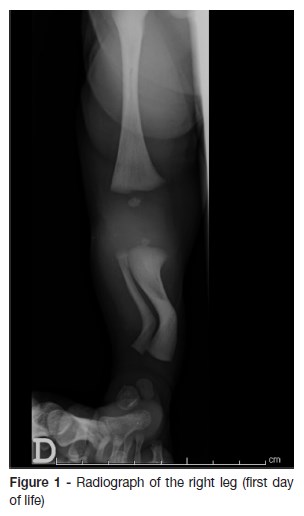
A male newborn, from a medically supervised pregnancy, had obstetric ultrasounds showing a hypoplasia of the right leg bones and ipsilateral deviation of the foot. He was born at 38 weeks gestation by vaginal delivery. On physical examination at birth, a medial deviation of the inferior third of the right leg and dorsiflexion of the right foot was noticed, with no other findings. The right leg x-ray showed a posteromedial bowing of the tibia (figure 1).
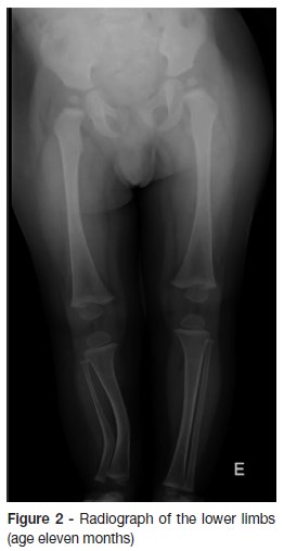
He had an excellent outcome with a conservative approach (figure 2).
What is your Diagnosis?
DIAGNOSIS
Congenital posteromedial bowing of the tibia (CPMBT)
DISCUSSION
Congenital deformities of the tibia are characterized by a bowing of the tibial diaphysis. According to the direction of the apex of the deformity, they can be classified in anterolateral, anteromedial and posteromedial.1-3
CPMBT is characterized by a calcaneal valgus deformity and is the most benign of all the congenital deformities of the tibia.1-5 If there is a large angulation of the diaphysis, the deformity is usually obvious at birth.1-3 However, if there is only a slight angulation, only a thorough examination of the lower limbs can identify it.1 CPMBT is distinguishable from the anterolateral bowing due to the absence of pseudoarthrosis or association with type I Neurofibromatosis (featured in 50% of the latter) and distinguishable from the anteromedial bowing because it is not associated with the absence of fibula or lateral segments of the foot.1,4
The etiology of this deformity has not yet been clarified, however three hypotheses have been proposed: an abnormal positioning in the uterus; circulatory changes; embrionary changes.1,4
Usually CPMBT resolves spontaneously until the age of eight. Limb length discrepancy is the main complication. To avoid it, clinical and imagological surveillance is recommended until the skeletal maturity.1,6
A conservative approach is the first line in most cases. However, the choice of treatment varies with the degree of limb length discrepancy, age, target height and family/patient preference.4
This case pretends to enlighten the rarity of this skeletal deformities and its possible association with other diseases. It is fundamental that Neonatologists/Pediatricians are aware of these pathologies to assure an early and correct referral.
REFERÊNCIAS BIBLIOGRÁFICAS
1. Dias AIM, Pinheiro L, Almeida E. Deformidade póstero-medial congénita da tíbia: a propósito de 2 casos clínicos. Nascer e Crescer. 2013; 22:171-3. [ Links ]
2. Kaufman SD, Fagg JA, Jones S, Bell MJ, Saleh M, Fernandes JA. Limb lengthening in congenital posteromedial bow of the tibia. Strat Traum Limb Recon. 2012; 7:147–53. [ Links ]
3. Ferguson J, Wainwright A. Tibial bowing in children. Orthopaedics And Trauma. 2012; 27:30-41. [ Links ]
4. McCarthy J. Tibial bowing. Emedicine.medscape.com. Abril, 2015. (Accessed 5 june 2017). Available at: http://emedicine.medscape.com. [ Links ]
5. Shah H, Rousset M, Canavese F. Congenital pseudarthrosis of the tibia: Management and complications. Indian J Orthop. 2012; 4:616-26. [ Links ]
6. Shah HH, Doddabasappa SN, Joseph B. Congenital posteromedial bowing of the tibia: a retrospective analysis of growth abnormalities in the leg. J Pediatr Orthop B 2009; 18:120-8. [ Links ]
CORRESPONDENCE TO
Ivete Afonso
Department of Pediatrics
Hospital Pedro Hispano
Unidade Local de Saúde de Matosinhos
Rua Dr. Eduardo Torres
4464-513 Senhora da Hora, Matosinhos
Email: ivete.afonso@hotmail.com
Received for publication: 14.06.2017 Accepted in revised form: 23.10.2017

